Review Report
Assisted reproductive technologies in cervids: current status and emerging needs
Jamie Stewart
Virginia-Maryland College of Veterinary Medicine, Virginia Polytechnic Institute and State University, Blacksburg, VA, USA
Abstract
With over 40 cervid species in the world, the use of assisted reproductive technologies to enhance genetic selection in farmed deer operations can appear somewhat complicated. In the US alone, various species of cervids are farmed captively, with the industry focused on selecting genetic traits (e.g. carcass growth and antler conformation) in males to be sold for trophy hunting. Due to these unique aspects of the cervid industry, assisted reproductive technologies are frequently used to disseminate valuable genetics and increase profitability for breeding farms. Compared to domesticated ruminants, few studies have thoroughly evaluated semen freezing procedures, hormonal synchronization, AI techniques and embryo technologies within each individual cervid species. Veterinarians, therefore, rely heavily on extrapolated and anecdotal information when making decisions aimed at improving reproductive success in farmed cervid species. This review describes the published data that is heavily focused on white-tailed and red deer and discusses anecdotal evidence that is commonly used within the industry. An analysis of gaps in knowledge is also included to help determine what techniques may be developed in the future that could benefit cervid farmers.
Keywords: Cervid, deer, embryo, reproduction, semen
Citation: Clinical Theriogenology 2026, 18, 14657, http://dx.doi.org/10.58292/CT.v18.14657
Copyright: © 2026 The Author(s). This is an Open Access article distributed under the terms of the Creative Commons Attribution-NonCommercial 4.0 International License (http://creativecommons.org/licenses/by-nc/4.0/), permitting all noncommercial use, distribution, and reproduction in any medium, provided the original work is properly cited.
Published: 13 November 2026
CONTACT: Jamie Stewart jlstewart13@vt.edu
Competing interests and funding: None to declare.
Introduction
Captive deer farming is a continuously growing industry in North America, with trophy hunting and venison products commonly used as the end market. White-tailed deer (Odocoileus virginianus) is the most popular cervid species raised in the US; others include wapiti/elk (Cervus canadensis), red deer (Cervus elaphus), mule deer (Odocoileus hemonionus), fallow deer (Dama dama), sika deer (Cervus nippon), and axis deer (Axis axis). Reindeer (Rangifer tarandus). another popular cervid species raised in the US, are more commonly used for exhibition purposes. In general, progress for the development of assisted reproductive technologies (ARTs) in reindeer has lagged behind that of other farmed deer, due to lesser demand.1 As per a 2017 report,2 white-tailed deer farming generated ~ $7.9 billion, compared to $3 billion in 2007.3 Breeding stock represents the major component of this industry (> $900 million in 2017) as it involves all the animals raised and sold.2
With the focus of cervid farming in the US being on the production of male offspring for trophy hunting, males are frequently selected for larger carcass size and broad, well-branched antlers.4 As with other ruminants, artificial insemination (AI) with frozen or fresh semen is the most effective tool available to select for and disseminate these desirable traits.4 The high demand for producing more terminal bucks per season has led to increased interest in technologies that can alter offspring sex proportion, including sex-sorted semen, laparoscopic artificial insemination (LAI), and multiple ovulation embryo transfer (MOET). These technologies rely heavily on effective semen cryopreservation to preserve and disseminate genetics from valuable males. This article provides a comprehensive review on the reproductive physiology of popular farmed cervid species and summarizes the current breeding technologies available for advancing the deer farming industry.
Reproductive physiology and seasonality
The Cervidae family contains over 40 species; that complicates breeding management because the reproductive physiology of 1 species cannot always simply be extrapolated into other species.5 Most of the species farmed in the US are of northern temperate origin and exhibit strict seasonality as short-day breeders. However, the most notable exception is the axis deer, a tropical species with an extended reproductive season.5 Similar to other small ruminant species, photoperiod is the main driver for the seasonal effects on reproduction in deer. As day length begins to shorten following the summer solstice, production of melatonin from the pineal gland increases. Melatonin has a stimulatory effect on the hypothalamus, driving an increase in gonadotropin releasing hormone (GnRH) pulsatility; GnRH acts on the anterior pituitary gland to trigger luteinizing hormone (LH) secretion that stimulates testosterone production from testes and is responsible for triggering ovulation. Interestingly, although rams and goat bucks tend to maintain the ability to produce semen outside the natural breeding season, cervid males mostly become completely infertile (except for the axis stag).
Within the reproductive season, females of most deer species are polyestrus and continue to cycle until they become pregnant or enter anestrus in the spring. Silent ovulations are common early in the breeding season, wherein the female will ovulate without exhibiting any signs of estrus. These ovulations are typically not fertile but will result a corpus luteum (CL) that frequently has a shortened lifespan. This process allows exposure of the hypothalamus to progesterone priming, increasing estradiol receptor expression in the hypothalamus and allowing for estrus expression with the subsequent ovulation. In addition to the progesterone priming that is necessary also for fertile ovulations, short cycles are also believed to promote synchronization within the herd.5
There are variations among species in estrous cycle lengths: 7-17 days in sika deer,6 17 days in axis deer,5 19-20 days in reindeer,7,8 18-20 days in red deer,5 20-23 in wapiti/elk,9 21-23 days in fallow deer,5 and 21-30 days in mule deer and white-tailed deer.10 Mean duration of the estrous cycle in most cervid species tends to increase throughout each breeding season.5 Estrous behavior tends to last up to 24 hours, but frequently will terminate in response to a successful copulation. This mechanism has been reported in a variety of spontaneously ovulating species, including cattle, sheep, goats and deer.11 Although initially considered to occur secondary to a mechanical neuroendocrine reflex,12-14 more recent studies have proposed that male-produced pheromones15 or factors in the seminal plasma16 may also have a role in hastening ovulation in ruminants in response to breeding. Regardless, the cessation of estrus and promotion of ovulation are advantageous for the male by limiting opportunities for other males to mate. Further work evaluating these breeding mechanisms in each cervid species could improve our ability to manipulate the reproductive cycle and enhance success of ARTs.
Semen collection and cryopreservation
The most effective and established way to disseminate valuable genetics in the cervid industry is through semen collection and cryopreservation. Most cervid males are highly seasonal and will only produce quality semen during rut. In white-tailed bucks, sperm motility and morphology both peak around December in the US (when there is hard antler), and start declining after the winter solstice.17 Serum testosterone concentrations have similar peaks, with ~ 88% of males measuring > 80 ng/ml, whereas 100% of males measured < 20 ng/ml by March (when the antlers have shed).17 Consequently, the quality of frozen-thawed white-tailed deer semen was significantly greater for ejaculates collected in December versus those collected from the same bucks in September or March.18 Similar patterns have also been reported in other cervid species (e.g. red deer,19 wapiti,20 fallow deer,21 sika deer,22 and reindeer23). On the contrary, Axis deer raised in south Texas had bigger testis and total epididymal sperm content in the summer compared to other species and that is when the majority of stags were also in hard antler.24 In contrast to temperate species, Axis deer stags maintained testicular function throughout the year, regardless of antler status.24 As expected, however, testicular volume, serum testosterone concentration, semen volume, sperm concentration, percentage of morphologically normal sperm, and percentage of motile sperm were all greater when Axis deer stags were in hard antler.25
Semen collection procedures in cervids typically require complete immobilization of the male to safely and effectively facilitate electroejaculation.26 In tamer animals (e.g. reindeer, trained red deer stags) an artificial vagina, modified for deer, can also be utilized to collect semen, similar to other ruminants.1,27 Electroejaculation has also been performed while restrained without anesthesia in wapiti28 and Sika deer.22 For electroejaculation, immobilization can be performed in a variety of ways. Butorphanol-azaperone-medetomidine injectable combination in white-tailed deer bucks provided an effective plane of anesthesia with no adverse outcomes following semen collection.29 Another injectable anesthetic that the author has frequently used for semen collection in white-tailed deer is a combination of tiletamine-zolazepam and xylazine.17,18,30 If the animal’s temperament and working facilities allow, hand injection of anesthetics is preferred to minimize risks associated with darting via projector (dart gun) that can include tissue bruising or necrosis, bone fractures, broken needles, or inadvertent penetration of the thoracic or abdominal cavities. Throughout rut, when male cervids are in hard antler, there are inherent risks to both the handlers and animals when using working facilities unless the antlers have already been removed; therefore, many practitioners elect to give tranquilizers remotely with a dart gun. Alternatively, a pole syringe may be an option in tamer animals that can be safely approached but should still be done from outside a holding facility.26 Immobilization with an intravenous or intramuscular injection of xylazine and ketamine has also been used successfully for performing electroejaculation in red deer,19,31 fallow deer,32 white-tailed and mule deer33 and Axis deer.25 Medetomidine with ketamine can also be used for anesthesia.33 Alpha-2-agonists (xylazine and medetomidine) can be reversed using injectable tolazoline, yohimbine or atipamezole to hasten recovery after the procedure is completed.
Most extenders used for ruminant sperm cryopreservation can be used successfully in cervid species. Commercial egg yolk, soybean and liposome-based semen extenders were all effective for cryopreservation of white-tailed buck semen in a prior study, with the soybean-based extender producing sperm with greater progressive motility on postthaw analysis.30 Similarly, the same soybean-based extender was acceptable for freezing red deer semen.31 In author’s experience, most protocols used to cryopreserve domestic ruminant semen are adequate for cervid semen.34 Both soybean and liposome-based extenders are popular with practitioners because they only require having distilled water (rather than needing to acquire and process fresh eggs), and semen can be frozen in a 1-step method.
Cervid semen is commonly collected on farm and then transported for processing. A small aliquot of ejaculate should be evaluated immediately at low magnification using light microscopy to ensure adequate concentration and viability. The sample should then be diluted at a ~ 1:1-1:3 ratio of semen to prewarmed (37°C) extender, depending on the perceived concentration. To begin preparation for processing, it is helpful to place both the tube with the extended sample and any excess extender in the warm water bath, but unplug it during transit to the laboratory, enabling both to slowly start cooling to room temperature. Once ready for processing, the concentration of the extended sample should be measured, followed by dilution to the desired final concentration (usually ~ 100-200 x 106 sperm/ml in a 0.25 or 0.5 ml straw). Semen cryopreservation protocols should always be refined in a way that works best for each facility. Semen can be loaded into straws at room temperature before placing in a cold room (4°C) or loaded while in a cold room after the equilibration period (usually 3-5 hours). If loading straws at room temperature, it is important to bundle the straws together into a 65 mm goblet (maximum 150 (0.5 ml) straws per goblet) to store in the cold room to ensure that they do not cool too quickly. After the desired equilibration period (depends on the extender), the straws can be frozen. Although automated semen freezing units can be used, straws can also simply be placed on a rack in a Styrofoam box 4-6 cm above the surface of liquid nitrogen for 10-15 minutes, then submerged into the liquid and loaded into canes for storage, as done in other species.
Epididymal sperm harvesting is another procedure that can be easily extrapolated from techniques performed in domesticated ruminants. Deer farmers may wish to submit testes from males that either died unexpectedly or males that they hunted and decided that they wanted to preserve the genetics. Clients should be instructed to remove the testes with as much of the ductus deferens as possible, seal them in a clean plastic bag, cover the bag in a towel, and send to the clinic on ice within 24 hours. Testes should be evaluated to ensure that they are an appropriate size for flushing (Figure 1A).35 The cauda epididymis can then be excised from its attachment to the body of the epididymis and testes (Figure 1B). From there, 2 methods are possible. In the first method, the ductus deferens can be uncoiled from the base of the epididymis as much as possible (Figure 1C). A blunted 25-G needle can then be placed into the free end of the ductus deferens and extender slowly infused until it begins to drip from the incised edge of the epididymis into a tube. If this method is unsuccessful (unable to flush or there are leaks), the entire epididymis and ductus deferens can be placed in a petri dish, excised with a scalpel blade, and flushed with extender. From there, the semen can be processed as described above, noting that a higher percentage of morphological defects, namely distal cytoplasmic droplets, may be present, but are unlikely to affect fertility if viability is good.
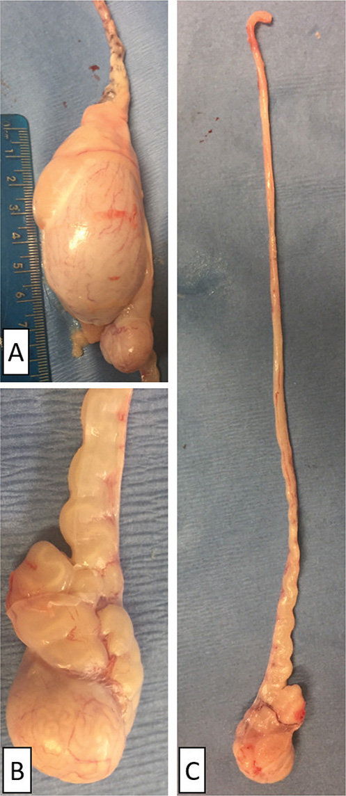
Figure 1. Preparation for epididymal flushing: evaluate and measure the testis prior to flushing (A); examine the ductus deferens lumen for distention with semen (B); carefully dissect entire cauda epididymis and associated ductus deferens from testis and epididymal body (C)
Estrus synchronization and timed artificial insemination
Synchronization protocols for timed artificial insemination are relatively similar amongst cervid species, with some noteworthy modifications. A controlled internal drug release (CIDR) insert or progestin-embedded sponge is commonly placed intravaginally at the start of estrus synchronization. The Eazi-breed CIDR contains progesterone embedded in a molded silicone insert that is absorbed transvaginally for up to 21 days and prevents females from ovulating. Sheep CIDR insert (contains 0.3 g of progesterone) is used for red deer, sika deer, fallow deer, reindeer and white-tailed deer, whereas cattle CIDR insert (contains 1.38 g of progesterone) is used in the larger bodied wapiti/elk.36 The CIDR is removed after 12-14 days in most cervid species, except for red deer, in which they are removed after 10-12 days.36 An injection of pregnant mare serum gonadotropin/equine chorionic gonadotropin (PMSG/eCG) is then usually given at CIDR removal to augment endogenous gonadotropins to promote selection and growth of recruited follicles directly from the ovary via follicle stimulating hormone (FSH) like activity.37,38 This FSH activity will also accelerate granulosa cell proliferation, increasing estrogen synthesis from the developing follicles that will eventually trigger the LH surge and promote a synchronized ovulation.37-39 Approximate doses (in IU units) of PMSG are: 50 in sika deer, 100-200 in white-tailed deer, 150-200 in red deer, and 190-200 in wapiti/elk.36 PMSG is omitted in fallow deer due to variable responses. The AI procedure is then scheduled to be performed at ~ 60 hours after CIDR removal. GnRH (~ 1-2 ml) may also be given at AI to promote ovulation, with some practitioners preferring to only give GnRH if PMSG is not used in the synchronization protocol.
Artificial insemination
Artificial insemination is commonly performed intravaginally, transcervically or laparoscopically in does/hinds. Estrus synchronization is usually recommended, as it can be challenging to perform estrus detection effectively in most cervid species. Electronic estrus detection system was useful for detecting estrus in red deer hinds subjected to transcervical or intracervical AI following a hormonally induced estrus or a spontaneous estrus.40 The pregnancy rates were, however, lower with transcervical/intracervical AI based on estrus (29-42%)40 than those reported for timed LAI. Although most producers can perform transvaginal or intracervical AI themselves, more advanced insemination techniques (e.g. transcervical and laparoscopic intrauterine) have better success rates and usually require trained personnel, further necessitating the need for estrus synchronization and timed procedures.
Transcervical AI (TCAI) is a method wherein the AI insemination gun is passed through the cervix to dispense semen into the uterine body. This procedure can be performed either while the female is anesthetized (Figure 2A) or while standing in a chute designed for cervids (Figure 2B). In smaller-bodied cervids (e.g. white-tailed deer) this procedure is usually performed using a clean vaginal speculum to visualize the external os of the cervix and then passing the insemination rod into the cervix and blindly manipulating the rod through the cartilaginous cervical rings until the uterine body is penetrated; after which semen is deposited slowly. In some females, it may not be possible to pass the rod through all the cervical rings and, therefore, the semen can be slowly deposited intracervical. It is beneficial to withdraw the speculum partially before depositing the semen to allow the vaginal wall to collapse over the end of the insemination rod and prevent backflow of semen while inseminating.41 Similar to cattle, in larger-bodied cervid species (e.g. wapiti/elk), transrectal palpation can be utilized to more effectively facilitate TCAI.
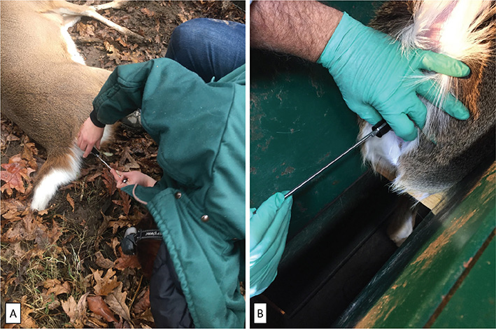
Figure 2. Transcervical AI in white-tailed deer: if no handling equipment is available, this technique (A) can be performed in an immobilized doe; nonanesthetized doe can be restrained in a deer handling chute (B), an ideal way
LAI is commonly performed in the cervid industry when utilizing valuable frozen-thawed semen, due to greater success rates compared to intravaginal and transcervical/intracervical techniques. Furthermore, LAI enables efficient semen utilization, since only a small dose of semen is needed compared to other techniques and multiple females can be serviced with a single straw. Additionally, the procedure is also more advanced, with most states in US requiring a veterinary license to perform or, at minimum, a veterinarian on site during the procedure. Due to the time required to set up equipment, female synchronization is necessary so that an entire group can be inseminated during the same visit. Females must be immobilized; that can be performed using injectable anesthetics (as described above for semen collection). Hand injection of females in a deer-handling chute is preferable over darting to minimize the risk of complications and ensure safe immobilization.
Once anesthetized, the caudal abdomen is clipped up to the site of the umbilicus and hair removed with a shop vacuum. A surgical preparation of the abdomen should then be performed. A local block with 2% lidocaine infused intramuscularly into each of the 2 port sites is sometimes performed to reduce incision site pain following the procedure. The 2 port sites are generally identified as ~ 6-8 cm cranial to the udder and 6-8 cm lateral to midline. After placing the doe in a Trendelenburg position, with the hind legs tightly secured, an incision can be made into the skin and underlying fascia at each port site. Some practitioners prefer to insert the insufflation needle (or teat canula) and infuse CO2 into the peritoneum first, while watching to ensure that the abdomen is distending equally, to minimize the risk of perforating abnormal organs with the trocar. Other practitioners prefer to insert a trocar first and confirm that the insufflation needle is not in any abdominal organs (e.g. urinary bladder, rumen, or intestine) prior to insufflating. Most practitioners prefer a smaller diameter (5 mm) trocar, but up to 10 mm trocars can be used. Once the abdominal cavity has been insufflated and viewed using the laparoscope through the first port site (Figure 3A), a second trocar can be placed at the contralateral port site. The semen should be loaded into a specialized AI sheath that contains a needle and infused into the greater curvature of 1 or both uterine horns (Figure 3B). Semen is likely to diffuse to both horns following infusion into a single horn; however, some practitioners prefer to infuse both horns either due to technical reasons (e.g. needing to infuse > 1 straw due to owner preference and/or semen quality) or to ensure that at least 1 injection is successful. Following successful insemination, maximize CO2 recovery from the abdomen and close port sites with staples or sutures. Reversal of anesthesia is recommended to hasten recovery.
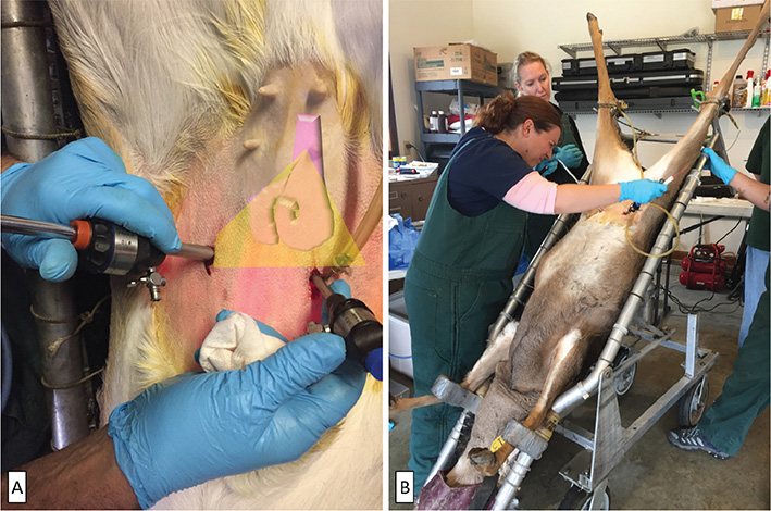
Figure 3. Laparoscopic AI in a white-tailed doe; first port site (left) is typically used to view the uterus with the laparoscope (A). After visualization of uterus, a second trocar is inserted into the right port site (B); insemination gun is then placed through the second port site so that semen can be infused into the greater curvature of 1 or both uterine horns
There are limited published data available on acceptable semen doses for various AI techniques among cervid species. Both intravaginal and intracervical deposition of semen (~ 20-80 x 106 motile sperm) in red and fallow deer produced < 40% conception rates that increased to ~ 60-70% by adding a second insemination ~ 24 hours after the first insemination in red deer and increasing the dose of sperm used in both red and fallow deer to ~ 100 x 106 motile sperm.42 TCAI versus LAI with 0.25 ml frozen-thawed semen straws in white-tailed deer had no difference in fawning rates (40% for TCAI versus 45% for LAI). Similarly, when frozen-thawed semen (0.5 ml containing 10 x 106 progressively motile sperm before freezing) was deposited into the cervix or uterus of a synchronized white-tailed deer doe, pregnancy success was 17 times more likely than those receiving intravaginal insemination.43 In general, the pregnancy rates reported in white-tailed deer studies from both TCAI and LAI were lower than other reports of ~ 60-70% with TCAI and 75-85% for LAI.4 The author frequently freezes white-tailed deer sperm at a concentration of 120 x 106 sperm per ml in 0.5 ml straws to ensure that the minimum recommended dose of 12-15 x 106 progressively motile sperm (postthaw) for performing TCAI is achieved.44 On the contrary, only 6-8 x 10⁶ progressively motile sperm (postthaw) is recommended for LAI to be successful. It is, therefore, not uncommon for owners to request that 0.5 ml straws be divided among 2 females when performing LAI (which seems to produce acceptable results).45 Further studies that evaluate minimum effective semen doses for laparoscopic versus TCAI are needed for each deer species to develop standardized breeding dosages for semen freezing. Additionally, studies that evaluate how ‘splitting straws’ affects pregnancy rates would also be useful to provide practitioners with tools to use when discussing the implementation of these procedures with deer farmers.
Artificial insemination using sex-sorted semen
In small ruminants, sex-sorted semen is feasible due to the satisfactory results reported with low doses when laparoscopically inseminated directly into the uterus.46,47 In contrast to the domesticated ruminants, where selection of females for production of milk and replacement dams is valued, the farmed deer industry would benefit more from achieving a higher proportion of male offspring to be sold for trophy hunting.
White-tailed deer have an advantage over domestic animals with excellent semen quality that has strong tolerance to cryopreservation when collected during rut.17,18,30 Sex-sorted white-tail deer semen using flow cytometry increased postthaw motilities and decreased DNA fragmentation when compared to conventional cryopreserved semen.48 Similarly, there was no difference in postthaw motility of control, X or Y sorted semen collected from Sika deer; however, a greater percentage of intact acrosomes were observed in sorted groups.49 Anecdotally, pregnancy rates of 93-95% have been reported using cryopreserved sex-sorted cervine doses of 8- 9 x 10⁶ sperm.50 With fresh, sex-sorted sperm, doses of 4 x 10⁶ sperm have also reportedly produced ~ 5-8% better conception rates than conventional semen in deer.50 The pregnancy rates of Sika hinds undergoing LAI were greater with unsorted frozen-thawed sperm (~ 79%, 0.25 ml straws containing 2.8 x 10⁶ sperm per straw) than for those inseminated with either X (~ 46%) or Y sorted (~ 48%) sperm (0.25 ml straws containing 2.3 x 10⁶ sperm per straw).49 A recent case publication reported 67-80% conception rates using fresh, Y-sorted semen (~ 4.5-5 x 10⁶ sperm) inseminated laparoscopically in synchronized white-tailed deer, producing 85-100% male offspring.51 In a large, controlled study using red deer (Cervus elaphus), pregnancy rates were greater for conventional frozen-thawed semen (~ 78%) versus Y sex sorted (50%) when inseminated transcervically.52 Additional in vivo studies are needed to improve the success rates amongst deer species, as sex-sorted semen has so far proved to be successful in the minimal reports available. LAI is likely the most feasible technique to ensure greater pregnancy success, but studies evaluating dose-dependent outcomes of sex-sorted semen with both laparoscopic and TCAI are still needed to assist practitioners with making evidence-based recommendations.
Multiple ovulation and embryo transfer
White-tailed deer have been at the forefront of ARTs in the US, whereas ARTs in red deer have been studied more extensively in other countries. MOET techniques have been adapted in many deer species from the surgical techniques developed for sheep and goats. Preparation of donor deer for MOET is similar to that described in other ruminants; progesterone devices (e.g. CIDRs or intravaginal sponges containing medroxyprogesterone acetate) are placed for 12-14 days and FSH injections administered twice daily beginning 3-4 days before removal of progesterone devices.44,53 The recommended dosage of FSH can vary based on species, protocol, product and individual animal response, with some practitioners using tapering doses, whereas others prefer a consistent dose throughout the protocol. Folltropin (Vetoquinol), a porcine pituitary derived source of FSH, is the only commercially available FSH product in the US and is approved for cattle. Recombinant FSH products have much promise for the cervid industry and have become commercially available in many countries outside of the US.4 Within the US, compounded recombinant FSH use has been discussed but is strongly discouraged by the food animal residue avoidance database as it does not fulfill the necessary requirements for treatment under the animal medicinal drug use clarification act.54 In red deer, a typical protocol begins with insertion of a progesterone device (day 0) for 12 days, with FSH treatment twice daily (for a total of 8 doses) from days 10-13, and removing the progesterone device between 12-24 hours prior to final FSH dose.55 GnRH agonist (buserelin acetate) can also be given 12 hours after the final FSH dose to stimulate and synchronize ovulations.53 Fallow deer protocols are similar except that progesterone devices are used for 14 days, with 8 doses of FSH twice daily beginning on days 10.5-11, with the last dose at progesterone device removal.42,56 Intramuscular PMSG (100 IU) is also given either at the first or last FSH injections in fallow deer protocols.55,56 Similar white-tailed deer protocols have been described, with a CIDR for 12-14 days and twice daily FSH treatment for 4 days, beginning 3 days prior to CIDR removal.44 Following superovulation, breeding can be performed by turning out males for natural mating or performing AI as described above.
Embryo recovery in all cervid species occurs ~ 8-9 days after progesterone withdrawal and is frequently performed surgically, similar to sheep and goats. In larger-bodied cervid species (e.g. wapiti/elk) nonsurgical embryo collection techniques performed transcervically, similar to cattle, have been successful.57 For surgical embryo recovery, animals should be anesthetized using combinations (e.g. tiletamine-zolazepam and ketamine) or a combination of an alpha-2-agonist (e.g. xylazine or medetomidine) and ketamine. Inhalant anesthesia is strongly recommended to maintain anesthesia throughout the procedure but may not be available in a field setting, so additional injectable doses of anesthetics should be readily available if needed. Butorphanol-azaperone-medetomidine is not recommended for this procedure as it does not provide adequate anesthesia for a surgical procedure.
When an adequate plane of anesthesia is achieved, the female should be placed in dorsal recumbency in a cradle with the abdomen clipped and surgically prepped from the umbilicus caudally. Antibiotics and antiinflammatories are necessary during preparation to minimize the risk of infection and decrease postoperative pain/inflammation. A local block is also useful to desensitize the incision site. After draping the abdomen, the female is then positioned ~ 30 degrees in a Trendelenburg position. Many practitioners choose to evaluate the ovaries via laparoscopy first to avoid performing surgery on a female that did not respond to superovulation. The procedure is performed similarly to that described for LAI, with the focus being on visualization of multiple CL on the ovaries. A ventral midline laparotomy is then performed such that the uterus can be easily exteriorized. Some reports have stated that 0.5 mg of clenbuterol given IV can reduce uterine turgidity to improve ease of uterine exteriorization; however, this is not legal in food producing animals in the US.54,55 A foley catheter is placed into the base of the first uterine horn after creating a small hole with closed hemostats. The balloon can then be inflated using flush media or air to seal the base of the uterine horn. A tom cat catheter or 18 G intravenous catheter is placed into the lumen at the tip of the uterine horn, near the uterotubal junction. The uterine horn is then flushed in a retrograde manner from the uterotubal junction to the base of the uterine horn using ~ 50-60 ml of embryo flush media into a petri dish or embryo filter. The procedure is repeated in the second uterine horn. Uterine incisions can be closed using 1 or 2 inverting stitches. The body wall and skin are closed as with any routine abdominal incision, and reversal of anesthesia is recommended when needed to hasten recovery. Embryos can be transferred fresh (within a few hours) or cryopreserved.
Embryo recovery rates average ~ 5.5 embryos per white-tailed doe44 and 3.1 embryos per red deer hind.53 Embryo grading and staging should be performed as in other domesticated ruminants.58 Additionally, fresh embryo handling and embryo cryopreservation protocols used in cervids have been largely extrapolated from validated techniques used in cattle. Slow-freezing methods using glycerol as a cryoprotectant were used historically, but have fallen out of favor due to multistep procedures needed for thawing. More recent protocols have evaluated slow freezing with ethylene glycol and vitrification for cervid embryo cryopreservation.53 Pregnancy rates in red deer hinds were ~ 64% for fresh embryos, 53% for vitrified/warmed embryos and 70% for ethylene glycol frozen/thawed embryos following surgical transfer.53 Vitrified embryos were also successfully used for cryopreservation in sika deer, resulting in pregnancy rates of 62% for vitrified embryos versus 86% for fresh embryos and birth rates of 54% for vitrified embryos versus 64% for fresh embryos.59 Reported implantation rates in white-tailed deer does are ~ 90% for fresh embryos and ~ 65% for frozen embryos.44,53
Similar to embryo recovery, embryo transfer in cervids is also frequently performed surgically, with the main exception being wapiti/elk, due to their larger stature. As in other domesticated ruminants, recipient synchronization is necessary. Estrus synchronization for deer embryo transfer recipients is similar to protocols for AI, with 12-14 days of progesterone exposure (frequently through insertion and removal of intravaginal inserts), with or without PMSG treatment at progesterone withdrawal. In fallow deer, progesterone withdrawal is recommended to occur in recipients 24 hours prior to that in donors to ensure adequate synchronization, and no PMSG is recommended, due to highly variable results.55,56 Recipients should be anesthetized and surgically prepared, as described for donors. Laparoscopic examination of the ovaries is recommended prior to laparotomy to confirm females have at least 1 CL. Uterine horn ipsilateral to the CL(s) should then be exteriorized for embryo transfer. This technique can be performed entirely surgically through a ventral midline incision (similar to that described for flushing) or laparoscopically assisted, where the laparoscope is used to exteriorize the tip of the uterine horn through an extension of the port site where the transfer will be performed. One or 2 embryos are typically deposited into the uterine horn using a tomcat catheter.
Further work to refine MOET protocols in farmed cervid species should improve success rates and increase dissemination of valuable female genetics. Most notably is the need to refine superovulation protocols and increase the number of embryos generated per female. Additionally, exploring new ways to perform embryo flushing and transfer nonsurgically may improve female longevity within breeding programs and increase practicality of MOET in cervid species. One older report in white-tailed deer details a nonsurgical embryo collection technique in which a modified 16 G Verres needle was used to catheterize the cervix in 3 donor animals but only yielded 1 morula per female (ranging from poor to excellent quality).60 Nonsurgical embryo recovery protocols have been developed and refined in goats over the past decade with much success, using techniques that could be extrapolated to deer. Such techniques require a vaginal speculum to properly visualize the cervix, forceps to manipulate the external cervical os and uterine dilators to transpose the cervical rings and allow for a catheter to be introduced transcervically.61 Similar techniques may be beneficial in deer, as they could be performed in an appropriate deer handling facility while under sedation rather than under general anesthesia.
In vitro produced embryos
Although some work with in vitro produced (IVP) embryos is being performed in cervids, this technology is not widely available in white-tailed deer farmed in the US. Vitrification of oocytes has been described in domesticated ruminant species, but there is limited research that has assessed this procedure in cervids.62 Rather, oocyte collection that can be performed both surgically and laparoscopically is used to harvest oocytes specifically for in vitro embryo production, as embryo cryopreservation is much more successful than oocyte vitrification.62 In a recent study done in the US, IVP embryos were successfully generated from white-tailed deer using oocytes acquired surgically.63 An average of 6.3 viable oocytes were obtained from each donor.63 An IVP embryo protocol that included both in vitro oocyte maturation and in vitro fertilization was adapted from commercial bovine protocols, resulting in a cleavage rate of 62% and a blastocyst rate of 44%.63 Similarly, oocytes collected surgically from captive white-tailed deer does had an average of 6.3 viable oocytes per donor.64 Embryos were also generated successfully using IVP procedures extrapolated from beef cow protocols that included maturation for 24 hours and fertilization for 18-20 hours with media designed for small ruminants, resulting in a cleavage rate of 40% and a blastocyst rate of 37%.64 Embryos were then incubated for 96 hours, evaluated for grading and staging, and then vitrified.64 Oocyte collection and IVP procedures have also been developed and described in red deer62,65 and sika deer,66 with an emphasized application on both management of farmed deer and extrapolation to endangered deer species. Oocyte maturation was performed for 23 hours in 5% CO2 humidified air (~ 20% oxygen) in red deer, followed by coincubation with sperm for 15 hours for in vitro fertilization.67 In another protocol for deer embryos, oocyte maturation is performed over 22-24 hours, in vitro fertilization is performed over 18 hours and embryo culture is performed for 4 days, after which visual assessment every 24 hours is performed to either transfer or freeze, based grading and staging.65 There are apparently no reported pregnancy rates for IVP embryos in white-tailed deer but in red deer, pregnancy rates have ranged from 30-63%.62 Using frozen IVP embryos prepared from abattoir-derived red deer cumulous oocyte complexes, pregnancies were achieved in both red deer (29%) and wapiti recipients (45%) after transcervical transfer.68 However, further work is needed to refine these techniques for species farmed in the US and determine if, from a cost perspective, IVP embryos would be beneficial over the more established AI and MOET procedures for enhancing genetic selection in captive deer farms.
Compared to MOET (requires synchronization and superovulation of females to be successful), OPU techniques do not require as much handling prior to performing the oocyte retrieval. In domesticated ruminants, estrus synchronization and FSH priming are sometimes performed prior to OPU, though their efficacy in increasing oocyte production and quality is questionable. For wild cervids, FSH priming may not be practical, due to the need to perform serial injections over several days. In a study, white-tailed does were synchronized prior to oocyte retrieval with a single dose of FSH at CIDR removal;63 however, no synchronization was described prior to oocyte retrieval in another IVP study involving white-tailed does,64 despite generating the same number of viable oocytes.63,64 There was, however, a greater cleavage and blastocyst rate in the study where synchronization was performed.63,64 Future studies that directly compare hormonal stimulation prior to OPU in each cervid species are indicated to best determine whether synchronization and superovulation would improve oocyte yield and embryo production per donor.
Another benefit of using IVP embryos over MOET in cervids is the ability to perform a less-invasive laparoscopic OPU technique to retrieve oocytes, as described in sheep and goats,69 red deer70 and sika deer.71 Although nonsurgical embryo collections were successful in smaller-bodied cervid species,60 surgical techniques still appear to be widely preferred due to the difficulty in passing a catheter through the cartilaginous cervix.72 Although both MOET and IVP embryo techniques can also utilize sexed semen, in vitro fertilization would require fewer viable, sex-sorted semen than MOET. Embryo collection and transfer was successfully performed in wapiti/elk and the number of transferable embryos per hind did not differ between sex-sorted Y sperm and unsorted semen.57 Research on evaluation of sex-sorted semen for IVP embryos in deer species is lacking, so no meaningful comparison of sex-sorted semen with MOET can be made. Although it appears that many ARTs can be extrapolated from other ruminant species, more studies are needed to best refine these procedures, with an emphasis on developing less invasive modifications. With further refinement, commercialization of these procedures could be implemented in the future for the deer industry to enhance genetic selection.
Pregnancy diagnosis
The capstone of any good reproductive program is to verify pregnancy. Since most deer species have a limited breeding season, owners are typically anxious to turn the females out with males quickly after any breeding attempts to ensure that pregnancy occurs within the breeding season. When evaluating AI success, many owners elect to simply assess lengths of pregnancy during the fawning/calving season to estimate AI pregnancy rates. It is, therefore, beneficial that owners are aware of species-specific lengths of pregnancy amongst cervids: 183-210 (average ~ 200) days for white-tailed deer/mule deer,73 198-221 (average ~ 211) days for reindeer,74 228-234 days for fallow deer,42,75 229-237 (average ~234) days for red deer42,76 and 240-252 (average ~ 247) days for wapiti/elk.77 The wide range in pregnancy length can make pregnancy rates unreliable if there was male exposure shortly after AI or ET. Owners utilizing this method for designating the sire should consider DNA testing fawns/calves to confirm results, especially if they are being sold.
For any female undergoing surgery as a donor or recipient, a minimum resting period of 2 weeks after the procedure is recommended to allow for healing prior to turn out with a male for clean-up breeding. For recipient females, it would be valuable to test for pregnancy before making the decision to turn out with a male to prevent any added stress that could compromise pregnancy. The quickest and most reliable pregnancy detection technique is transrectal ultrasonography ~ 30-60 days postAI,77 performed relatively easily within a deer-handling chute using a transducer handle. The differences between day 30 pregnancy and days 50-60 pregnancy should be obvious based on placentomes and fetal skeleton formation in the latter (Figure 4) and can be used to differentiate AI pregnancies from natural cover. A blood test that quantifies serum pregnancy-specific protein B can also be used in most ruminant species after ~ day 25 of pregnancy as this protein is produced by binucleate cells in the ruminant placenta.78 However, compared to ultrasonography, the blood test cannot differentiate between various stages of pregnancy, predict fetal number, or assess fetal viability.78 An animal-side test has recently been released (Pregnostx On-Site Pregnancy Test Kit for Cattle, Sheep and Goats, BioTracking Inc.) that may make it practical for owners with a deer handling chute to perform pregnancy diagnosis, provided that they can collect blood. This on-site test kit was developed in the same lab that validated the pregnancy-specific protein B laboratory test (BioBryn, BioTracking, Inc.), but there are few studies available that have validated its use in ruminants as of yet.79
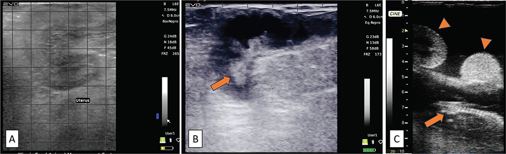
Figure 4. Transrectal ultrasonographic images of deer uterus A. appearance of a nonpregnant uterine horn; B. distended uterine horns with anechoic fluid and an embryo (arrow) visible on day 30; C. fetal skeleton can be easily visualized (arrow) along with several placentomes (arrowhead) on day 60
Conclusion
The implementation of ARTs in captive deer farms does not need to be a daunting task for most practitioners. Many of the techniques that have been described herein have been extrapolated from those developed in cattle, sheep and goats. Practitioners who have established semen-freezing protocols for domesticated ruminants should find it to be a very similar process in deer. The biggest difference that may require a learning curve is understanding safe handling procedures and immobilization protocols. Compared to domesticated ruminants, deer species also synchronize easily using the deer-specific protocols described herein. Depending on the species, success rates in performing TCAI and LAI are relatively high, with the biggest difference from small ruminants and cattle also being restraint and handling. Sex-sorted semen technologies may also enable producers with enhancing genetic selection and increasing profitability and should be discussed when planning for the upcoming breeding season.
Although more advanced reproductive procedures (e.g. MOET, IVP) are typically reserved for valuable animals, continued growth in the farmed deer industry may necessitate further advancement of these procedures. There are limited continuing education opportunities available to assist practitioners with learning how to perform embryo flushing or OPU in cervids. However, the procedure is performed similarly in sheep and goats (for which there are ample learning opportunities) and can easily be extrapolated for practitioners proficient in those species or those willing to learn and practice. Lastly, if asked to work with deer species, practitioners should become well versed with state-specific legislation regarding captive deer farming and work with their state veterinarian to become accredited to perform tuberculosis and brucellosis testing, write certificate of veterinary inspections and assist producers with chronic wasting disease herd certification programs. Being well rounded in all aspects of deer farming and able to perform a variety of deer-specific veterinary services will be necessary and invaluable to deer farming clients.
References
| 1. | Lindeberg H, Nikitkina E, Nagy S, et al: Potential applications of assisted reproductive technologies (ART) in reindeer (Rangifer tarandus). Anim Reprod Sci 2021;235:1-10. doi: 10.1016/j.anireprosci.2021.106890 |
| 2. | Anderson DP, Outlaw JL, Earle ML, et al: Economic Impact of the US Deer Breeding and Hunting Operations. College Station, TX: Agricultural and Food Policy Center, Texas A&M University; 2017. |
| 3. | Anderson DP, Frosch BJ, Outlaw JL: Economic Impact of the United States Cervid Farming Industry. College Station, TX; Agricultural and Food Policy Center, Texas A&M University: 2007. |
| 4. | Bringans M: The captive deer industry in the USA and Mexico: Details on breeding, nutrition, and handling. In: John F: editor. The Management of Enclosed and Domesticated Deer: International Husbandry Systems and Diseases. Springer Nature Switzerland AG; Cham, Switzerland: 2022. p. 267-282. |
| 5. | Asher GW: Reproductive cycles of deer. Anim Reprod Sci 2011;124:170-175. doi: 10.1016/J.ANIREPROSCI.2010.08.026 |
| 6. | Higuchi N, Ohnishi N, Minami M: Temporal pattern of estrus in free-ranging sika deer (Cervus nippon). Mammal Study 2013;38:275-279. doi: 10.3106/041.038.0402 |
| 7. | Ropstad E, Forsberg M, Sire JE, et al: Plasma concentrations of progesterone, oestradiol, LH and 15-ketodihydro-PGF2α in Norwegian semi-domestic reindeer (Rangifer tarandus tarandus) during their first reproductive season. Reproduction 1995;105:307-314. doi: 10.1530/JRF.0.1050307 |
| 8. | Ropstad E: Reproduction in female reindeer. Anim Reprod Sci 2000;60-61:561-570. doi: 10.1016/S0378-4320(00)00100-7 |
| 9. | McCorkell R, Woodbury M, Adams GP: Ovarian follicular and luteal dynamics in wapiti during the estrous cycle. Theriogenology 2006;65:540-556. doi: 10.1016/J.THERIOGENOLOGY.2005.05.049 |
| 10. | Knox WM, Miller KV, Marchinton RL: Recurrent estrous cycles in white-tailed deer. J Mammal 1988;69:384-386. doi: 10.2307/1381395 |
| 11. | Morales-Piñeyrúa JT, Ungerfeld R: Pampas deer (Ozotoceros bezoarticus) courtship and mating behavior. Acta Vet Scand 2012;54:60. doi: 10.1186/1751-0147-54-60 |
| 12. | Zarrow MX, Clark JH: Ovulation following vaginal stimulation in a spontaneous ovulator and its implications. J Endocrinol 1968;40:343-352. doi: 10.1677/JOE.0.0400343 |
| 13. | Romano JE, Alkar A, Amstalden M: Effect of copulation on estrus duration and ovulation time in goats. Theriogenology 2016;85:330-334. doi: 10.1016/j.theriogenology.2015.09.021 |
| 14. | Romano JE, Benech A: Effect of service and vaginal-cervical anesthesia on estrus duration in dairy goats. Theriogenology 1996;45:691-696. doi: 10.1016/0093-691X(95)00415-5 |
| 15. | Parsons SD, Hunter GL: Effect of the ram on duration of oestrus in the ewe. J Reprod Fertil 1967;14:61-70. doi: 10.1530/jrf.0.0140061 |
| 16. | Stewart JL, Stella S, Cunha LL, et al: Administration of nerve growth factor-β to heifers with a pre-ovulatory follicle enhanced luteal formation and function and promoted LH release. Theriogenology 2020;148:37-47. doi: 10.1016/j.theriogenology.2020.02.040 |
| 17. | Stewart JL, Shipley CF, Ellerbrock RE, et al: Physiological variations in reproductive and metabolic features of white-tailed deer (Odocoileus virginianus) bucks throughout the rutting season. Theriogenology 2018;114:308-316. doi: 10.1016/J.THERIOGENOLOGY.2018.04.015 |
| 18. | Stewart JL, Shipley CF, Ellerbrock RE, et al: Variation in post-thaw sperm quality of white-tailed deer bucks (Odocoileus virginianus) during rut. Anim Reprod Sci 2018;195:121-130. doi: 10.1016/J.ANIREPROSCI.2018.05.014 |
| 19. | Garde JJ, Martínez-Pastor F, Gomendio M, et al: The application of reproductive technologies to natural populations of red deer. Reprod Domest Anim 2006;41:93-102. doi: 10.1111/J.1439-0531.2006.00773.X;WGROUP:STRING:PUBLICATION |
| 20. | Haigh JC, Cates WF, Glover GJ, et al: Relationships between seasonal changes in serum testosterone concentrations, scrotal circumference and sperm morphology of male wapiti (Cervus elaphus). J Reprod Fertil 1984;70:413-418. doi: 10.1530/JRF.0.0700413 |
| 21. | Gosch B, Fischer K: Seasonal changes of testis volume and sperm quality in adult fallow deer (Dama dama) and their relationship to the antler cycle. J Reprod Fertil 1989;85:7-17. doi: 10.1530/JRF.0.0850007 |
| 22. | Cheng FP, Wu JT, Chan JPW, et al: The effect of different extenders on post-thaw sperm survival, acrosomal integrity and longevity in cryopreserved semen of Formosan Sika deer and Formosan Sambar deer. Theriogenology 2004;61:1605-1616. doi: 10.1016/J.THERIOGENOLOGY.2003.07.015 |
| 23. | Nagy S, Lindeberg H, Nikitkina E, et al: Reproduction of male reindeer (Rangifer tarandus). Anim Reprod Sci 2021;227:106722. doi: 10.1016/J.ANIREPROSCI.2021.106722 |
| 24. | Willard ST, Randel RD: Testicular morphology and sperm content relative to age, antler status and season in axis deer stags (Axis axis). Small Rumin Res 2002;45:51-60. doi: 10.1016/S0921-4488(02)00084-6 |
| 25. | Umapathy G, Sontakke SD, Reddy A, et al: Seasonal variations in semen characteristics, semen cryopreservation, estrus synchronization, and successful artificial insemination in the spotted deer (Axis axis). Theriogenology 2007;67:1371-1378. doi: 10.1016/J.THERIOGENOLOGY.2007.01.019 |
| 26. | Boesch JM: Anesthesia of white-tailed deer and other cervids. Clinical Theriogenology 2011;3:511-515. Available from: https://clinicaltheriogenology.net/index.php/CT/article/view/10155 [cited 1 June 2026]. |
| 27. | Giżejewski Z: Effect of season on characteristics of red deer /Cervus elaphus L./ semen collected using modified artificial vagina. Reprod Biol 2004;4:51-66. |
| 28. | Haigh J, Barth A, Cates W, et al: Electro-ejaculation and semen evaluation of wapiti. In: Biology of Deer Production. Proceedings of the International Conference Held at Dunedin, New Zealand. 1985: p. 197-203. Available from: https://www.cabidigitallibrary.org/doi/full/10.5555/19850191909 [cited 7 June 2026]. |
| 29. | Kirschner SM, Rodenkirch R: Assessment of butorphanol-azaperone-medetomidine combination as anesthesia for semen collection and evaluation of semen quality in white-tailed deer (Odocoileus virginianus). Anim Reprod Sci 2017;184:196-203. doi: 10.1016/J.ANIREPROSCI.2017.07.016 |
| 30. | Stewart JL, Shipley CF, Katich AS, et al: Cryopreservation of white-tailed deer (Odocoileus virginianus) semen using soybean-, liposome-, and egg yolk-based extenders. Anim Reprod Sci 2016;171:7-16. doi: 10.1016/J.ANIREPROSCI.2016.05.006 |
| 31. | Martínez-Pastor F, Martínez F, Álvarez M, et al: Cryopreservation of Iberian red deer (Cervus elaphus hispanicus) spermatozoa obtained by electroejaculation. Theriogenology 2009;71:628-638. doi: 10.1016/j.theriogenology.2008.09.033 |
| 32. | Asher GW, Kraemer DC, Magyar SJ, et al: Intrauterine insemination of farmed fallow deer (Dama dama) with frozen-thawed semen via laparoscopy. Theriogenology 1990;34:569-577. doi: 10.1016/0093-691X(90)90012-I |
| 33. | Shipley CF: Breeding soundness examination of the male ovine, caprine and cervidae species. Clinical Theriogenology 2016;8:445-448. |
| 34. | Asher GW, Berg DK, Evans G: Storage of semen and artificial insemination in deer. Anim Reprod Sci 2000;62:195-211. doi: 10.1016/S0378-4320(00)00159-7 |
| 35. | Sugai N, Shipley CF, Ellerbrock RE, et al: Testicular size as an indicator of poor sperm output in white-tailed (Odocoileus virginianus) and mule (Odocoileus hemionus) deer bucks. Clinical Theriogenology 2018;10:358-358. doi: 10.58292/CT.V10.10066 |
| 36. | Tamassia M: Synchronization of estrus of whitetail deer and other select cervids, sheep & goats. Clinical Theriogenology 2011;3:531-535. Available from: https://clinicaltheriogenology.net/index.php/CT/article/view/10159/16143 [cited 30 April 2026]. |
| 37. | Liu J, Zhao M, Zhuang S, et al: Low concentrations of o,p’-DDT inhibit gene expression and prostaglandin synthesis by estrogen receptor-independent mechanism in rat ovarian cells. PLoS One 2012;7:e49916. doi: 10.1371/JOURNAL.PONE.0049916 |
| 38. | Choi JK, He X: Improved oocyte isolation and embryonic development of outbred deer mice. Sci Rep 2015;5:12232. doi: 10.1038/srep12232 |
| 39. | Wang C, Liu X, Shang S, et al: Hormone guided estrus synchronization using progesterone sponge and PMSG in goats: a cost-effective optimization strategy. Front Vet Sci 2026;13:1743340. doi: 10.3389/FVETS.2026.1743340/TEXT |
| 40. | Bowers SD, Brown CG, Strauch TA, et al: Artificial insemination following observational versus electronic methods of estrus detection in red deer hinds (Cervus elephus). Theriogenology 2004;62:652-663. doi: 10.1016/J.THERIOGENOLOGY.2003.11.019 |
| 41. | Shipley CF: Equipment and techniques for transcervical artificial insemination and laparoscopic insemination in small ruminants. Clinical Theriogenology 2011;3:525-530. |
| 42. | Asher GW, Fisher MW, Fennessy PF, et al: Oestrous synchronization, semen collection and artificial insemination of farmed red deer (Cervus elaphus) and fallow deer (Dama dama). Anim Reprod Sci 1993;33:241-265. doi: 10.1016/0378-4320(93)90118-B |
| 43. | Gentry GT, Lambe J, Forbes W, et al: The effect of equine chorionic gonadotropin (eCG) on pregnancy rates of white-tailed deer following fixed-timed artificial insemination. Theriogenology 2012;77:1894-1899. doi: 10.1016/J.THERIOGENOLOGY.2012.01.007 |
| 44. | Coe J, Bringans M: Physiology and reproductive techniques for the whitetail deer. Clinical Theriogenology 2020;12:268-270. Available from: https://clinicaltheriogenology.net/index.php/CT/article/view/9241 [cited 30 April 2026]. |
| 45. | Kensell K: Semen handling for insemination in the field: splitting straws, loading the equipment and juggling eight straws at a time. Clinical Theriogenology 2011;3:523-524. Available from: https://clinicaltheriogenology.net/index.php/CT/article/view/10157 [cited 30 April 2026]. |
| 46. | de Graaf SP, Beilby KH, Underwood SL, et al: Sperm sexing in sheep and cattle: the exception and the rule. Theriogenology 2009;71:89-97. doi: 10.1016/J.THERIOGENOLOGY.2008.09.014 |
| 47. | González-Marín C: Evolution of cervine, caprine and ovine sex-sorted semen processing. Clinical Theriogenology 2018;10:205-209. doi: 10.58292/CT.V10.9914 |
| 48. | Kjelland ME, González-Marín C, Gosálvez J, et al: DNA fragmentation kinetics and postthaw motility of flow cytometric-sorted white-tailed deer sperm. J Anim Sci 2011;89:3996-4006. doi: 10.2527/JAS.2011-4014 |
| 49. | Gao QH, Wei HJ, Luo J, et al: Flow cytometric sexing of X- and Y-chromosome-bearing sperm in Sika deer (Cervus nippon). Small Rumin Res 2009;81:100-104. doi: 10.1016/J.SMALLRUMRES.2008.11.014 |
| 50. | González-Marín C, Góngora CE, Moreno JF, et al: Small ruminant SexedULTRATM sperm sex-sorting: status report and recent developments. Theriogenology 2021;162:67-73. doi: 10.1016/J.THERIOGENOLOGY.2020.12.028 |
| 51. | Sugai N, Shipley C, Rothrock-Gensler L, et al: Laparoscopic artificial insemination of white-tailed deer with fresh sex-sorted semen. Clinical Theriogenology 2024;16:1-5. doi: 10.58292/CT.V16.10513 |
| 52. | Anel-López L, Garcia-Álvarez O, Tarantini T, et al: Influence of insemination time on the fertility of sex sorted frozen-thawed Y-sperm in red deer. Theriogenology 2018;113:171-175. doi: 10.1016/J.THERIOGENOLOGY.2018.03.005 |
| 53. | Soler JP, Mucci N, Kaiser GG, et al: Multiple ovulation and embryo transfer with fresh, frozen and vitrified red deer (Cervus elaphus) embryos in Argentina. Anim Reprod Sci 2007;102:322-327. doi: 10.1016/j.anireprosci.2007.03.024 |
| 54. | Clapham MO, Martin KL, Davis JL, et al: Extralabel drug use in wildlife and game animals. J Am Vet Med Assoc 2019;255:555-568. doi: 10.2460/JAVMA.255.5.555 |
| 55. | Fennessy PF, Asher GW, Beatson NS, et al: Embryo transfer in deer. Theriogenology 1994;41:133-138. doi: 10.1016/S0093-691X(05)80058-5 |
| 56. | Morrow CJ, Asher GW, Berg DK, et al: Embryo transfer in fallow deer (Dama dama): superovulation, embryo recovery and laparoscopic transfer of fresh and cryopreserved embryos. Theriogenology 1994;42:579-590. doi: 10.1016/0093-691X(94)90375-S |
| 57. | Gao QH, Wang HE, Zeng WB, et al: Embryo transfer and sex determination following superovulated hinds inseminated with frozen–thawed sex-sorted Y sperm or unsorted semen in Wapiti (Cervus elaphus songaricus). Anim Reprod Sci 2011;126:245-250. doi: 10.1016/J.ANIREPROSCI.2011.05.006 |
| 58. | Bó GA, Mapletoft RJ, Bó GA, et al: Evaluation and classification of bovine embryos. Anim Reprod 2018;10:344-348. Available from: http://www.animal-reproduction.org/article/5b5a604cf7783717068b46a2 [cited 19 November 2024]. |
| 59. | Wang L, Zhou GB, Shi WQ, et al: First live offspring born in superovulated sika deer (Cervus nippon) after embryo vitrification. Theriogenology 2012;78:1627-1632. doi: 10.1016/J.THERIOGENOLOGY.2012.07.008 |
| 60. | Magyar SJ, Hodges C, Seager SW, et al: Successful nonsurgical embryo collection with surgical transfer in captive white-tailed deer. Theriogenology 1988;29:273. |
| 61. | Fonseca JF, Zambrini FN, Alvim GP, et al: Embryo production and recovery in goats by non-surgical transcervical technique. Small Rumin Res 2013;111:96-99. doi: 10.1016/J.SMALLRUMRES.2012.08.007 |
| 62. | Korzekwa AJ, Kotlarczyk AM: Artificial reproductive technology (ART) applied to female cervids adapted from domestic ruminants. Animals 2021;11:2933. doi: 10.3390/ANI11102933 |
| 63. | Bangert E, Shipley C, Rabel R, et al: 32 The efficiency of an adapted bovine IVF protocol to produce in vitro-derived embryos from oocytes collected via surgical ovum pickup from live white-tailed deer (Odocoileus virginianus) donors under captivity in central Illinois. Reprod Fertil Dev 2023;36:165-166. doi: 10.1071/RDV36N2AB32 |
| 64. | Maraboto M, Bautista M, Francisco J, et al: Successful in vitro embryo production with oocytes aspirated from live white-tailed deer (Odocoileus virginianus texanus) donors under captivity in northeast Mexico. J Ethol Anim Sci 2026;7:1-7. doi: 10.23880/JEASC-16000143 |
| 65. | Berg DK, Thongphakdee A: In vitro culture of deer embryos. Methods Mol Biol 2019;2006:191-207. doi: 10.1007/978-1-4939-9566-0_14/FIGURES/2 |
| 66. | Locatelli Y, Hendriks A, Vallet JC, et al: Assessment LOPU-IVF in Japanese sika deer (Cervus nippon nippon) and application to Vietnamese sika deer (Cervus nippon pseudaxis) a related subspecies threatened with extinction. Theriogenology 2012;78:2039-2049. doi: 10.1016/J.THERIOGENOLOGY.2012.07.025 |
| 67. | Korzekwa AJ, Kotlarczyk AM, Szczepańska AA, et al: Antioxidative potential of red deer embryos depends on reproductive stage of hind as a oocyte donor. Animals 2020;10:1190. doi: 10.3390/ANI10071190 |
| 68. | Beaumont SE, Berg MC, Strongman K, et al: 188 Direct-thaw trans-cervical transfer of red deer frozen in vitro blastocysts can result in pregnancies. Reprod Fertil Dev 2004;17:244-244. doi: 10.1071/RDV17N2AB188 |
| 69. | Wieczorek J, Koseniuk J, Skrzyszowska M, et al: L-OPU in goat and sheep - different variants of the oocyte recovery method. Animals 2020;10:658. doi: 10.3390/ANI10040658 |
| 70. | Comizzoli P, Mermillod P, Cognié Y, et al: Successful in vitro production of embryos in the red deer (Cervus elaphus) and the sika deer (Cervus nippon). Theriogenology 2001;55:649-659. doi: 10.1016/S0093-691X(01)00433-2 |
| 71. | Locatelli Y, Vallet JC, Huyghe FP, et al: Laparoscopic ovum pick-up and in vitro production of sika deer embryos: Effect of season and culture conditions. Theriogenology 2006;66:1334-1342. doi: 10.1016/J.THERIOGENOLOGY.2006.05.005 |
| 72. | Waldhalm SJ, Jacobson HA, Dhungel SK, et al: Embryo transfer in the white-tailed deer: a reproductive model for endangered deer species of the world. Theriogenology 1989;31:437-450. doi: 10.1016/0093-691X(89)90549-9 |
| 73. | Short C: Morphological development and aging of mule and white-tailed deer fetuses. J Wildl Manage 1970;34:383. doi: 10.2307/3799025 |
| 74. | Shipka MP, Rowell JE, Sousa MC: Steroid hormone secretion during the ovulatory cycle and pregnancy in farmed Alaskan reindeer. J Anim Sci 2007;85:944-951. doi: 10.2527/JAS.2006-589 |
| 75. | Birgersson B, Ekvall K: Early growth in male and female fallow deer fawns. Behav Ecol 1997;8:493-499. doi: 10.1093/BEHECO/8.5.493 |
| 76. | Scott IC, Asher GW, Archer JA, et al: The effect of conception date on gestation length of red deer (Cervus elaphus). Anim Reprod Sci 2008;109:206-217. doi: 10.1016/J.ANIREPROSCI.2007.11.025 |
| 77. | Haigh JC: The gestation length of wapiti (Cervus elaphus) revisited. Anim Reprod Sci 2001;65:89-93. doi: 10.1016/S0378-4320(00)00222-0 |
| 78. | Duquette JF, Belant JL, Beyer DE, et al: Comparison of pregnancy detection methods in live white-tailed deer. Wildl Soc Bull 2012;36:115-118. doi: 10.1002/WSB.115;CTYPE:STRING:JOURNAL |
| 79. | Hameed OA, Nawasreh AN, Hag ME, et al: Comparative evaluation of pregnancy diagnosis in sheep using On-site pregnancy test (Pregnostx) and ultrasonography. World J Adv Res Rev 2024;22:1927-1935. doi: 10.30574/WJARR.2024.22.2.1600 |